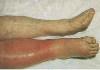
Deep Venous Thrombosis
Deep vein thrombosis (also called DVT) is a blood clot in a vein deep inside your body. These clots usually occur in one of the major deep veins of the lower legs, thighs, or pelvis. A clot blocks blood circulation through these veins, which carry blood from the lower body back to the heart. The blockage can cause pain, swelling, or warmth in the affected leg.your leg veins. While DVT is a fairly common condition, it is also a dangerous one. Occasionally the veins of the arm are affected (known as Paget-Schrötter disease). If tlood clot breaks away and travels through your bloodstream, it could block a blood vessel in your lungs. This blockage (called a pulmonary embolism) can be fatal. Statistics reveal that at least 650,000 patients die each year from pulmonary embolism, making it the third most common cause of death in the United States.

In the United States, about 2 million people per year develop DVT. Most of them are aged 40 years or older. Up to 600,000 are hospitalized each year for the condition.
View the following animation about DVT and Pulmonary embolus
Signs and symptoms
There may be no symptoms referrable to the location of the DVT ( 30-50% of the patients will experience no symptoms ), but the classical symptoms of DVT include pain, swelling and redness of the leg, dilation of the surface veins, gradual onset of pain, warmth to the touch, worsening leg pain when bending the foot, leg cramps, especially at night and/or bluish or whitish discoloration of skin.
A careful history has to be taken considering risk factors (see below ). On physical exam, several techniques can be used for the detection of DVT, such as measuring the circumference of the affected and the contralateral limb at a fixed point (to objectivate edema), and palpating the leg veins, which is often tender. Physical examination is unreliable for excluding the diagnosis of deep vein thrombosis.
Causes of DVT
Three factors may lead to formation of a clot inside a blood vessel.
1. Damage to the inside of a blood vessel due to trauma or other conditions
2. Changes in normal blood flow, including unusual turbulence, or partial or complete blockage of blood flow
3. Hypercoagulability, a rare state in which the blood is more likely than usual to clot ( your physician should order blood tests to diagnose a primary hypercoagulability state ).
Risk Factors of DVT
Prolonged sitting, such as during a long plane or car ride
Prolonged bed rest or lack of movement
Recent surgery, particularly orthopedic, gynecologic, or heart surgery
Recent trauma or fractured bones of the hip, thigh, or lower leg
Obese patients
Recent childbirth
High altitude ( Over 4000 masl )
Use of estrogen replacement (hormone therapy, or HT) or birth control pills
Cancer
Genetic changes in certain blood clotting factors
Disseminated intravascular coagulation (DIC)
Advanced age
A second DVT is much more likely to happen after a first one.
Diagnosis of DVT
Physical exam will consist of:
A. Homan’s test: Dorsiflexion of foot elicits pain in posterior calf. Rarely done due to its little diagnostic value and is theoretically dangerous because of the possibility of dislodgement of loose clot.
B. Pratt’s sign: Squeezing of posterior calf elicits pain.
These two medical signs do not perform well and are subjective. Lately clinical prediction rules that combine best findings are used to diagnose DVT. Scarvelis and Wells overviewed a set of clinical prediction rules for DVT in 2006.
Blood tests
Tests such as complete blood count, coagulation studies ( PT, APTT ), fibrinogen, liver enzymes, renal function and electrolytes are regularly ordered. Also a test know as D-dimer is performed. This cross-linked fibrin degradation product is an indication that thrombosis ( formation of a clot ) is occurring, and that the blood clot is being dissolved by plasmin. A negative D-dimer is definitive to exclude the possibility of a blood clot.
Imaging studies
The gold standard is intravenous venography, which involves injecting a peripheral vein of the affected limb with a contrast agent and taking X-rays, to reveal whether the venous supply has been obstructed. Because of its invasiveness, this test is rarely performed. Lately doppler ultrasonography which is a compression ultrasound scanning of the leg veins, combined with duplex measurements (to determine blood flow), can reveal a blood clot and its extent (i.e. whether it is below or above the knee). It has got high sensitivity, specificity and reproducibility.
Treatment
A. At home
 To increase comfort and lower the risk of the clot moving to the lung, the patient needs to keep the affected limb elevated, avoid prolonged sitting or bed rest and needs to use warm and moist heat to the area to relieve pain and inflammation.
Elastic compression stockings should be routinely applied beginning within 1 month of diagnosis of proximal DVT and continuing for a minimum of 1 year after diagnosis.
B. Medications
Anticoagulation is the usual treatment for DVT. In general, patients are initiated on a brief course (i.e., less than a week) of heparin ( fractionated or unfractionated ) treatment while they start on a 3- to 6-month course of warfarin.
C. Filter
If the patient can’t take warfarin, or if a blood thinner doesn’t work, your doctor may recommend that a filter is put into the vena cava (the main vein going back to your heart from your lower body). This filter can catch a clot as it moves through your bloodstream and prevent it from reaching your lungs. This treatment is used mostly for people who have had several blood clots travel to their lungs.
Hospital inpatient treatment is considered for patients with more than two of the following risk factors as these patients may have more risk of complications during treatment: bilateral DVT, renal insufficiency and cancer.


Leave a Reply